


Arthrosis (osteoarthrosis, deforming arthrosis) is a process of slow degeneration and destruction of the cartilage in the joint. The joint ends of the bones deform and grow, and the periarticular tissues become inflamed. The general diagnosis "arthrosis" means a group of diseases that are similar in symptoms but differ in origin. The joint - the affected area - consists of articular surfaces covered with cartilage tissue, a cavity with synovial fluid, a synovial membrane and a joint capsule. In advanced disease, it loses mobility and the patient experiences pain due to inflammatory processes.
reasons
Arthrosis of the joints develops due to the discrepancy between the amount of stress and the capabilities of the body. Lack of nutrients, excess body weight, heavy physical work and even sports can cause this.
Factors that influence the development of the disease:
- genetics, hereditary predisposition;
- age over 40 years;
- obesity, overweight;
- sedentary work, passive lifestyle;
- hard work, work that involves constant physical activity;
- inflammatory diseases;
- congenital joint pathologies (dysplasia);
- injuries, wounds;
- malfunction of the body (poor blood circulation, imbalance of hormones, trace elements).
The disease can be primary or secondary. The causes of primary arthrosis are still not well understood. Doctors believe that it develops in the presence of genetic factors (predisposition) and external adverse conditions.
Secondary arthrosis occurs against the background of inflammatory diseases, dysplasia and as a result of injuries, including professional ones.
Representatives of working professions and athletes have an increased chance of developing the disease. Representatives of the arts are also at risk: dancers (especially ballerinas), pianists. Arthrosis of the joints of the wrist and fingers most often affects people whose work involves fine motor skills: mechanics, mechanics and pianists. "Professional" arthrosis of loaders is localized in the knees, collarbones and elbows. Drivers, painters and miners suffer from elbow and shoulder joints. The weak point of ballerinas is the ankle. Athletes are also more likely to have injuries to the ankle and other joints of the handsand legs, depending on the type of sporting activity For example, a tennis player will be at high risk of shoulder and elbow joint disease.
Pathogenesis
Structural changes in cartilage occur due to an imbalance between tissue breakdown and repair. Collagen and proteoglycans are gradually "washed" from the body, new nutrients are not supplied. Cartilage tissue loses elasticity, becomes soft and does not withstand load.
Regardless of the location and the root cause, the disease develops in the same way. Gradually, the cartilage is completely destroyed, the ends of the bone "grind" against each other. The patient experiences pain, the intensity of which increases depending on the stage. The mobility of the joint gradually decreases, the patient's movements are limited.
p>Classification
Orthopedists use the classification formulated by the professor in 1961:
- Stage I. The bone becomes denser, the joint space narrows slightly. Discomfort during physical activity that disappears after rest;
- Stage II. The joint gap is significantly narrowed, the bone edges grow, the connective tissue becomes denser. The pain becomes constant, the muscles are hypertrophied, the joint is much less mobile, specific symptoms appear at the site;
- Stage III. The joint space is practically absent, the bony growths are large and destruction of the bone under the cartilage is possible. The joint is completely deformed and immobile. Acute or constant pain is possible depending on the type and location of the disease;
Depending on the location and form of the disease, symptoms, rate of development and treatment methods will vary.
Forms
The disease is characterized by a chronic form, but it can also appear in an acute form.
When the disease spreads to several joints (for example, fingers), it is called generalized.
Anatomical forms:
- deformation (osteoarthrosis). Leads to bony growths;
- uncovertebral. Destroys discs and intervertebral tissues in the cervical region;
- post-traumatic. It develops as a result of trauma, injury;
- rheumatoid. Autoimmune disease, inflammation of the connective tissue. It may be a consequence of previous arthritis;
- psoriatic. It develops against the background of psoriatic arthritis.
Localizations
Osteoarthritis is a disease that affects joints throughout the body.
spine. The causes can be autoimmune diseases, back diseases, increased stress, injuries, lack of trace elements, hormonal imbalance.
localizations:
- tail bone;
- lumbar region;
- thoracic vertebrae;
- cervical region
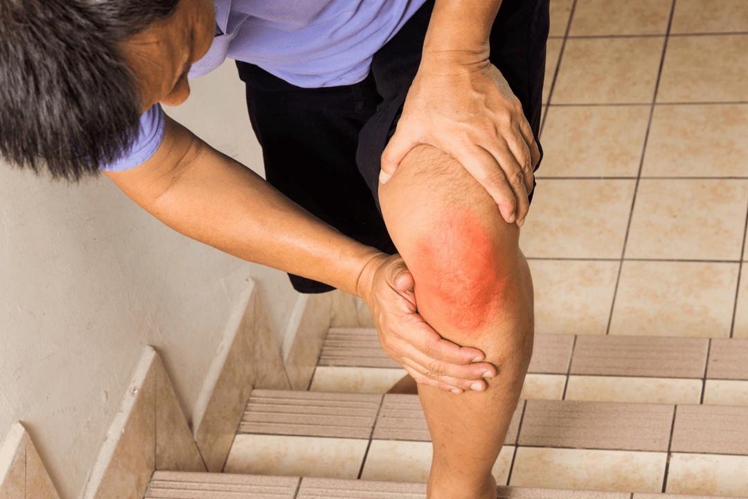
Legs. Knees and ankles are more prone to arthrosis. The reasons are injuries, excess weight, incorrect, excessive loads. Types of localization:
- gonarthrosis - knees;
- patellofemoral - femur and patella;
- ankle;
- talanovicular joint;
- feet and toes.
Hands. Lesions of the hands and fingers are more common and in most cases are related to occupational activity, injuries, age and hormonal changes. In addition, the disease is localized in the shoulder, wrist and elbow joints.
Torso. Localization in the body is less common compared to arthrosis of the limbs. Lesions are associated with professional activity, sedentary lifestyle (stagnation).
Types of localization:
- clavicle. When moving, "clicks" and pain are felt. At risk are athletes engaged in weight lifting and military personnel due to possible injuries;
- hip joints (coxarthrosis). The disease manifests itself as pain in the groin area.
Head>. Sometimes dental problems, vegetative disorders and even hearing loss are caused by damage to the temporomandibular joint. Edema disrupts the symmetry of the face, can affect the ear and cause headaches.
Symptoms
Symptoms of the disease depend on its location. Common manifestations for all species are:
- pain in the affected area. In the early stages - during movement, work, in the later stages - at rest;
- inflammation, swelling. Periarticular tissues swell, the skin becomes red;
- "clicking", crunching. Characteristic sounds are heard when moving;
- difficulty moving. As the disease progresses, the mobility of the affected area is impaired;
- reaction to cold. Many types of arthrosis are characterized by exacerbations in rainy and cold weather.
Exacerbations of the disease are associated with a general weakening of health. Due to viral diseases and increased stress, it takes an acute form and develops many times faster. During an exacerbation, symptoms, especially pain, become more pronounced. It is difficult for the patient to move to the point of complete loss of mobility and perform usual work.
Possible complications
The main danger is the loss of mobility of the joint, its deformation beyond the possibility of recovery. Due to the displacement of the axis, the pose is disturbed and the figure loses symmetry. Possible increased pressure on internal organs, their displacement, compression. Co-morbidities and malfunctions of body systems occur. For example, with coccyx arthrosis in women, gynecological complications are possible, and arthrosis of the temporomandibular joint or cervical spine causes disorders in the autonomic system: dizziness, sleep disorders. A patient with arthrosis can become disabled.
Diagnosis
To make a diagnosis, a comprehensive examination is carried out:
- history taking;
- radiography in several projections;
- MRI and CT to exclude tumors and obtain a three-dimensional image;
- blood and urine tests to rule out comorbidities and assess general health.
Depending on the cause of the disease, the patient is referred to a rheumatologist, traumatologist, surgeon or orthopedist.
Treatment
Stage I of the disease is best treated. Stage II patients can expect long-term relief from bone destruction. Stage III most often requires surgery.
Conservative (non-surgical) treatment:
- physiotherapy, use of orthoses, canes, crutches to reduce the load. Elimination of concomitant and aggravating factors (eg weight loss, stress, change of activity);
- taking nonsteroidal anti-inflammatory drugs. Selective COX-2 inhibitors are most effective. Chondroprotectors and atypical antidepressants are prescribed as auxiliary means;
- intra-articular injections of glucocorticoid hormones to reduce severe pain and inflammation.
Surgical methods:
- arthroscopy - internal examination of the joint and removal of cartilage fragments;
- arthroplasty - implantation of artificial cartilage;
- osteotomy - removal or dissection of bone tissue;
- chondroplasty - cartilage restoration;
- arthrodesis - artificial immobilization of a joint (most often ankle);
- endoprosthetics - removal and replacement of damaged joints with artificial ones.
Cardinal treatment allows you to stop the disease even at a late stage. Restoration of mobility is possible in individual cases (after replacement with an artificial one). However, this method is effective in the fight against pain. After the operation, it is necessary to recover with the help of physical therapy and medication methods.
Prognosis and prevention
After starting the treatment of I and II stage of arthrosis, there is a permanent improvement: pain and inflammation disappear. In this case, complete relief of the disease or its long-term preservation is possible.
In the treatment of arthrosis in stage III, improvements do not occur immediately. In some cases, the disappearance of pain is possible only after surgery. Often the joint remains immobilized or deformed. Patients with severe forms of arthrosis of the hip and knee joints receive group I or II disability.
It has been proven that there is no effective prophylaxis against arthrosis. Weight control, a balanced diet and moderate exercise will help reduce the risk of developing the disease. Examination at the first signs of arthrosis (especially after injuries and infectious diseases) and careful attention to health will allow you to identify the disease at an early stage.